Abdominoplasty
Breast augmentation
Breast reduction & uplift
Laser resurfacing
Fat Grafting
Liposuction
Melanoma
Sun protection
Basal cell cancer
Squamous cell skin cancers
Skin grafts
Facial repairs
Tissue expansion
Facial fractures
Trauma & burns


  The Pigment Cells (melanocytes) of the Skin & Dermoscopy  


Melanocytes
are found distributed in the skin

  • Scattered as individual cells at the junction of the upper (epidermis) and lower (dermis) layers of the skin. These cells produce granules of pigment (a suntan) in response to ultraviolet exposure.

  • Aggregated together (termed naevus cells) at various depths within the epidermis and dermis. Characteristic patterns of aggregation can usually be recognized by expert clinicians as those forming junctional, dermal, compound and dysplastic naevi (moles).

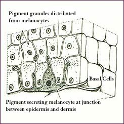
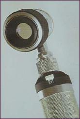
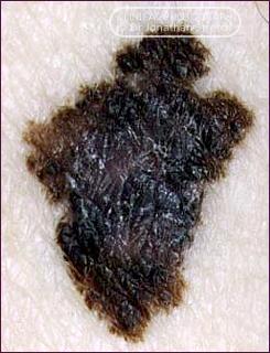
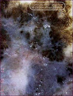
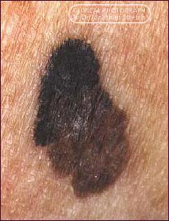
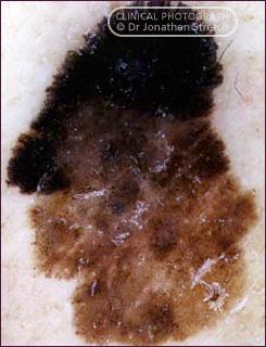
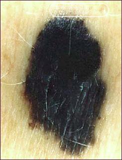
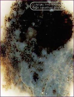
A number of changes in the pigment cell (melanocyte) network of the skin can be identified as potentially cancerous.

The Dermascope is an instrument which allows groups of pigment cells within the skin to be examined for changes which signal the development of a melanoma. It is a microscope which is used on living skin. Properly used, this instrument can substantially reduce the need to excise many naevi. Dermoscopy has greatly aided the diagnostic process.


Melanocyte at epidermo-dermal junction
Dermascope
 
   
Melanoma (Breslow thickness 2.1mm): Clinical photograph and dermascopic image
   
Melanoma (Breslow thickness 1.7mm): Clinical photograph and dermascopic image
   
Melanoma (Breslow thickness 2.4mm): Clinical photograph and dermascopic image


Jonathan Stretch Plastic Surgeon D.Phil (Oxon) F.R.A.C.S.